Where is the Carpal Tunnel Anatomically? In the Hand or in the Wrist?

Where is the Carpal Tunnel Anatomically? In the Hand or in the Wrist?


Question from George
Madison, Wisconsin
Answers From Doctors at First Hand Medical
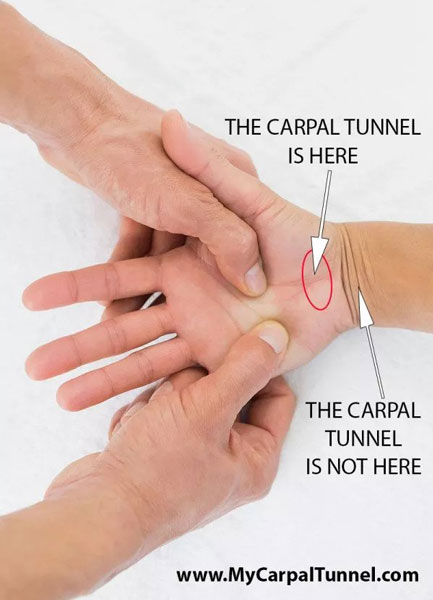
There is a lot of confusion about the anatomical location of the Carpal Tunnel among patients. Most patients think that it is in the wrist and associate wrist pain exclusively with Carpal Tunnel Syndrome. However, the Carpal Tunnel is not in the wrist at all. It is proximate to the wrist at the base of the hand between the two major muscle groups of the hand: The Thenar Muscles and the Hypothenar muscles. The photograph to the right identifies the exact anatomical location of the Carpal Tunnel between the two groups of major muscles at the base of the hand.
Three of the walls of the narrow passage known as the Carpal Tunnel are made of major bones supporting the structure of the hand. The fourth wall is the biggest ligament of the hand, known as the Transverse Carpal Ligament. This ligament connects the bones and Thenar Muscle Group and the Hypothenar Group of Muscles.
Grip strength in the hand is derived from the ligaments, bones and muscles are tightly working together to great a strong grip.
It is the Transverse Carpal Ligament that is severed in Carpal Tunnel Surgery. Surgeons hope the ligament heals back together with more space. When the ligament is severed in a carpal tunnel surgical procedure, the pressure on the Median Nerve in the Carpal Tunnel is relieved, because one of the four walls of the Carpal Tunnel has been compromised. Grip strength is also compromised by definition, this Transverse Carpal Ligament is integral to creating grip strength.
The Median Nerve and seven major tendons that control the movement in the hand from muscles in the forearm run through the Carpal Tunnel side by side.
Trapped inflammation and lymphatic fluid become toxic, viscous and block the normal healing processes of the hand.
The best way to relieve this blockage is actually through gentle consistent stretching action for eight hours each night during sleep over a six week healing period.
Applying Carpal Tunnel Surgery is akin to using a sledge hammer to repair fine cabinetry. There are better tools to use for such delicate work in tight spaces where there are so many important moving parts and are better left undisturbed. The body can heal itself if we give it the right natural treatment tools to unblock the healing pathways.
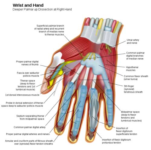
The expanded anatomical cross-sectional diagram of the hand taken from the Encyclopedia Britannica below shows another view of the Carpal Tunnel. You can see the Transverse Carpal Ligament and the Carpal Bones that make up the four walls of the Carpal Tunnel. You will also note how many important elements for proper hand function are packed into a tight space.
The Nerve Path of the Median Nerve is shown in the diagram to the left. The Median Nerve controls the Thenar Muscles and most of the minor muscles in the hand. When the nerve is pinched the neuromuscular sensations are restricted and muscle atrophy often occurs with Carpal Tunnel Syndrome. Wearing a wrist splint that restricts movement will aggravate and accelerate muscle atrophy of the hand overall and complicate the recovery from Carpal Tunnel Syndrome. This is why the Doctors at First Hand Medical recommend patients avoid rigid hand braces and wrist splints for the treatment of Carpal Tunnel Syndrome.
Natural healing should be the first line of treatment for all Carpal Tunnel Syndrome sufferers. Unleashing the powerful healing action of your body is the best way to get over Carpal Tunnel. Most people need help unblocking the lymphatic fluid flow and moving blocked inflammation.
Natural stretching during sleep is the best way to unblock these natural healing pathways.
 Created by renowned Harvard health care professionals.
